

UCLA Develops Palm-Held Portable Microscope For Healthcare Access
Imagine a palm-held portable, inexpensive microscope that serves remote areas by assisting doctors, nurses and field workers in improving healthcare worldwide.
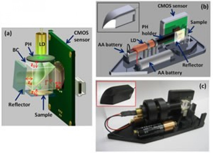
Researchers at the University of California (UCLA), Los Angeles, have actually built an on the go compact, light-weight, dual-mode microscope that is not only portable but also efficient. The prototype dual-mode microscope uses holograms instead of lenses as in the case of a traditional microscope.
Published in the Optical Society's (OSA) open-access journal, Biomedical Optics Express, the UCLA researchers explain that the prototype not only weighs the same as a medium-sized banana but also fits within the palm of a hand.
The researchers stated that since the microscope is based on parts which are mass-produced consumer electronics, the cost of the component materials in building the portable prototype could be anywhere between $50-$100 USD.
The prototype microscope with its two-in-one feature bears a transmission mode that can probe relatively large volumes of blood or water and a reflection mode that can image denser, opaque samples.
According to the paper, the spatial resolution for both modes is less than two micrometers which is comparable to that achieved by bulkier microscopes with low- to medium-power lenses. Aydogan Ozcan, senior author of the paper and an associate professor of electrical engineering and bioengineering, UCLA, said, This is the first demonstration of essentially a hand-held version of a microscope that can do dual-mode imaging within a very compact and cost-effective form.
Given minimal training, the device could be beneficial for doctors and field heath caregivers to improve healthcare in remote areas of the world which have little or no access to diagnostic equipment. Other benefits of the hand-held microscope could help ensure water quality, test patients' blood for harmful bacteria and even be used for semen-quality monitoring on animal farms.
It could also be useful for detecting outbreaks during health crises such as the recent outbreak of E. coli in Europe. Ozcan added, It's a very challenging task to detect E. coli in low concentrations in water and food. This microscope could be part of a solution for field investigation of water, or food, or maybe pathogens in blood.
An important advantage of the device is the light-weight model that enables users to replace the bulkier, heavier, more expensive pieces that most microscopes rely on for collecting and focusing light through the lenses.
The UCLA prototype uses the principle of hologram to recreate images from interfering light waves. Holograms are formed when light bouncing off (or passing through) a three-dimensional object is made to interfere with a reference beam, or light that has not hit the object.
An inexpensive light source is divided into two beams; one which interacts with microscopic cells or particles in the sample, and the other which does not interact at all. The beams then pass to an adjacent sensor chip, where their interference pattern is recorded.
The recording is fed into software which analyzes the pattern generated by recreating the path taken by the light that either passed through or bounced off the objects being imaged. The microscope thus captures raw data, but a computer is essentially required to reconstruct the images.
Field workers could make use of their laptops to process the information or send it over the Internet or mobile phone networks on to a remote server. Mobile phones could also be enabled to have sufficient processing power to carry out on-the-spot analysis. Ozcan noted, We are replacing an expensive and bulky, heavy component with computer codes.
Ozcan says, Each component of the device is fairly inexpensive. The laser light could come from a $5 laser pointer. The sensor chip that collects that light is the same as the ones in the backs of iPhones and Blackberrys and costs less than $15 per chip. And the whole image-collecting system runs on two AA batteries.
To achieve commercial distribution, Ozcan has set up a company to develop this technology to manufacture a version of the microscopes that could be sold to healthcare workers and hobbyists.
Ozcan says. Global health is a big field that requires better diagnostic tools, because resource-poor countries don't have the infrastructure for conducting essentially accurate diagnostic tests. There are so many problems that innovative solutions [like this microscope] would impact, he added
© Copyright IBTimes 2024. All rights reserved.
-
Eiffel Tower Loses Sparkle For Parisians Ahead Of Olympics
-

Former Number One Momota Retires From International Badminton At 29
-
Why Insurance Prices Have Skyrocketed
-
World Bank Aiming To Connect 250 Mn Africans To Energy Grid By 2030
-

IMF Says Global Debt Levels Face 'Great Election Year' Risk
-

Divisions Among Colombia's FARC Dissidents Complicate Peace Talks
-

French Far Right Gets Youthful Vibe With 28-year-old Leader
-

US Fed's Powell Says Inflation Fight May Take 'Longer Than Expected'
-
Mideast-related Oil Price Spike Threatens 'Relatively Good' Economic Outlook: IMF Chief Economist
-

Wine Growers 'On Tip Of Africa' Race To Adapt To Climate Change


