

Cancer-Killing Cells Caught in 3D at the Highest Resolution Ever [VIDEO & PHOTO]

Scientists have revealed more details on how white blood cells kill diseased tissue using deadly granules.
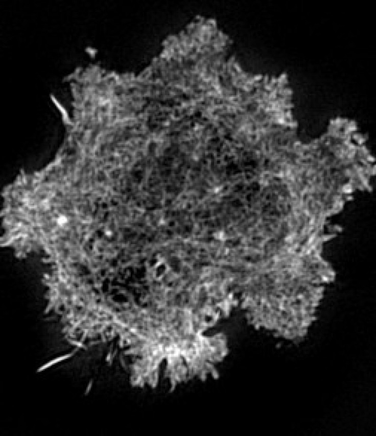
In a research published in PLoS Biology, researchers from Imperial College London and the University of Oxford, tell of how they used 'optical' laser tweezers and a super-resolution microscope to see the inner workings of white blood cells at the highest resolution ever.
They describe how a white blood cell reshuffles its scaffolding of actin proteins on the inside of its membrane in order to create a hole for delivering deadly enzyme-filled granules to kill diseased tissue.
For their study, the researchers looked at a type of white blood cell called a Natural Killer (NK) cell that offers the body protection by way of identifying and killing diseased tissue.
NK cells are important in our immune response to viruses and rogue tissues like tumors, said Professor Daniel Davis, from the Department of Life Sciences at Imperial College London, who led the research, in a statement. They may also play a role in the outcome of bone marrow transplants by determining whether a recipient's body rejects or accepts the donated tissue.
The researchers are hoping to learn more about how NK cells identify which tissues to kill and how it initiates the killing process. They believe this could lead to better health care for some patients.
In the future, drugs that influence where and when NK cells kill could be included in medical treatments, such as the targeted killing of tumors, Davis said. They may also prove useful in preventing the unwanted destruction by NK cells that may occur in transplant rejection or some auto-immune diseases.
Researchers were able to obtain the new visual resolution of NK cell in action as a result of a novel imaging technique created in collaboration with physicists at Imperial, and the use of a super high-resolution microscope at the University of Oxford, a press release stated.
The researchers were able to immobilize an NK cell and its target using a pair of 'optical' laser tweezers so that the microscope could capture all the action at the interface between the cells. They then observed inside the NK cell as the actin filaments separated to create a tiny portal. The enzyme-filled granules then moved to the portal, ready to pass out of the NK cell and onto the target to kill it.
The contact between an NK cell and its target is only about a hundredth of a millimeter across and the tiny actin proteins and granules change position continuously over the few minutes from initial contact until the target is killed, according to the press release.
These previously undetectable events inside cells have never been seen in such high resolution, said Dr. Alice Brown, also from the Department of Life Sciences at Imperial College London, in a statement. She conducted many of the experiments.
It is truly exciting to observe what happens when an NK cell springs into action, she added.
Watch a video of the cancer-killing cells caught in action below:
© Copyright IBTimes 2024. All rights reserved.
-

IMF Says Global Debt Levels Face 'Great Election Year' Risk
-

Divisions Among Colombia's FARC Dissidents Complicate Peace Talks
-

French Far Right Gets Youthful Vibe With 28-year-old Leader
-

US Fed's Powell Says Inflation Fight May Take 'Longer Than Expected'
-

Mideast-related Oil Price Spike Threatens 'Relatively Good' Economic Outlook: IMF Chief Economist
-

Wine Growers 'On Tip Of Africa' Race To Adapt To Climate Change
-

Despite Olympic Truce, Games Wrestle With Political Fallout
-

What Will The Fed Do With The Latest Inflation Numbers?
-

US Retail Sales Up More Than Expected In March
-

Alexandre De Moraes: Brazil Judge In Feud With Elon Musk


