

Ground-Breaking New Aqueous Reagent Turns Biological Tissue Transparent
A new reagent developed by researchers in Japan could turn biological tissues transparent, thereby offering an ideal means to analyze the complex organs and networks that sustain living systems.
The researchers at the RIKEN research organization in Japan claim that this advancement could take the process of medical imaging to new levels.
Experiments using fluorescence microscopy on samples treated with the reagent, published this week in Nature Neuroscience, have produced vivid 3D images of neurons and blood vessels deep inside the mouse brain.
Our current experiments are focused on the mouse brain, but applications are neither limited to mice, nor to the brain, states Atsushi Miyawaki, lead researcher at the RIKEN Brain Science Institute (BSI). We envision using Scale on other organs such as the heart, muscles and kidneys, and on tissues from primate and human biopsy samples.
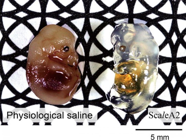
Scale, as the new chemical is called, overcomes all the limits of modern imaging processes by making the body tissues so clear that even light can pass deep enough to enable researchers get a clear picture of the underlying structures.
Most of the modern mechanical methods require that samples be sectioned into smaller pieces for visualization, while optical methods are prevented by the scattering property of light from probing deeper than 1mm into tissue.
Scale gets around these problems by doing two things together that no earlier technique has managed to do.
The first is to render biological tissue transparent. Scale does this significantly better than other clearing reagents and without altering the overall shape or proportions of the sample. The second is to avoid decreasing the intensity of signals emitted by genetically-encoded fluorescent proteins in the tissue, which are used as markers to label specific cell types.
This combination makes possible a revolution in optical imaging, enabling researchers to visualize fluorescently labeled brain samples at a depth of several millimeters and reconstruct neural networks at sub-cellular resolution.
© Copyright IBTimes 2024. All rights reserved.
-

French Air Traffic Controller Strike Threatens Flight Chaos
-

Azerbaijan Says 'Closer Than Ever' To Armenia Peace Deal Amid Border Talks
-

How UK's Biggest Water Supplier Sank Into Crisis
-

Taiwan Hit By Dozens Of Strong Aftershocks From Deadly Quake
-

Gaza Health System 'Completely Obliterated': UN Expert
-
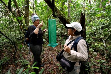
In Ecuadoran Amazon, Butterflies Provide A Gauge Of Climate Change
-

50 Years On, Vintage Vehicles To Reenact Portugal's Carnation Revolution
-

Conflicts Push Military Spending To 'All-time High': Report
-

'Thank You, America:' Zelensky And Netanyahu Applaud House Passage Of Foreign Aid Package
-

Women Journalists Bear The Brunt Of Cyberbullying


