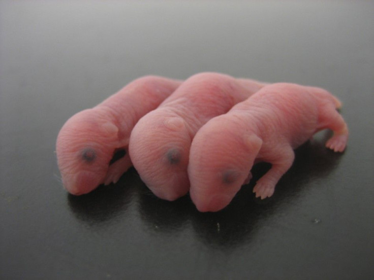
A Bit of Urine Turns Mouse Embryos Transparent

Japanese scientists have discovered that a little bit of solution containing urea can make mouse embryos transparent in just two weeks or less.
When scientists take photos of their mouse embryos, one of the biggest problems they face is the lack of transparency of the photos that prevent them from seeing deep inside tissues, according to geekosystem.com.
Scientists at RIKEN Brain Science Institute in Japan have found that a Scale solution can help researchers look inside tissue without making destructive insertions.
Even today's most promising techniques for visualizing biological tissue face this limitation: mechanical methods require that samples be sectioned into smaller pieces for visualization, while optical methods are prevented by the scattering property of light from probing deeper than 1mm into tissue. Either way, the full scope and detail of the biological sample is lost, RIKEN said in a press statement.
The Scale solution containing urea and glycerol allows researchers to examine the brain in 3D with the help of fluorescent markers. This recipe clears out tissue so well that the only limitation on how deep you can see is the power of the lens of the microscope, according to Discovery.com. And, as the researchers proved when they used it to image part of a mouse brain, it doesn't diminish the glow of the fluorescent tags.
Images drenched in the solution have revealed blood vessels deep within the embryo's brain at new depths and at sub-cellular resolution, the New Scientist reported.
In this experiment, the team of scientists only used the solution to study the brain but the potential of Scale goes much further. Atsushi Miyawaki, who led research, said, Our current experiments are focused on the mouse brain, but applications are neither limited to mice, nor to the brain.
We envision using Scale on other organs such as the heart, muscles and kidneys, and on tissues from primate and human biopsy samples, he added.
Looking ahead, Miyawaki's team has set its sights on an ambitious goal. We are currently investigating another, milder candidate reagent which would allow us to study live tissue in the same way, at somewhat lower levels of transparency. This would open the door to experiments that have simply never been possible before.
© Copyright IBTimes 2025. All rights reserved.


