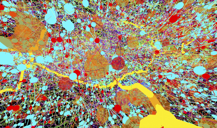
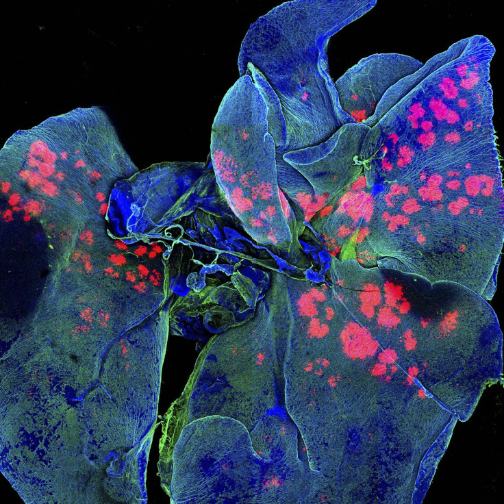
This is not abstract art, but a re-creation of the nervous system in a fruit fly larva.Albert Cardona, HHMI Janelia Research Campus, Wellcome Images
Science has a way of surprising even the most jaded observer, and that's certainly the case with the images selected for the Wellcome Image Awards 2015. The science images of the year feature cutting-edge technology and the hard work of researchers and their institutions. A panel of judges selected 20 images from the many acquired by the Wellcome Images library over the past year; the 20 will be honored at a March 18 ceremony, where an overall winner will be announced.
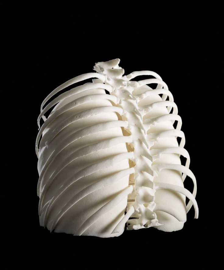
The 20 images cover a broad range of scientific topics, imaging techniques and materials. One photo from Dave Farnham depicts 3D-printed human lungs inside the rib cage of a woman with Hodgkin's lymphoma. The 3D rendering was created using data from her CT scans.
"The breathtaking riches of the imagery that science generates are so important in telling stories about research and helping us to understand often abstract concepts," Dr. Adam Rutherford, scientist and host for the Wellcome Image Awards 2015 ceremony, said in a statement. "It's not just about imaging the very small either, it's about understanding life, death, sex and disease: the cornerstones of drama and art. Once again, the Wellcome Image Awards celebrate all of this and more with this year’s incredible range of winning images."
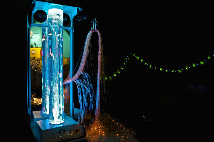
In addition to fascinating images of the human brain, boll weevils, pollen grains and a micrograph of a greenfly's eye, the photos include explore the human side of hospitals and research. Geraldine Thompson's image of a full pediatric sensory unit may look like a complicated mess of wires and lights meant for something complicated, but it's actually there to soothe young patients. The accompanying caption reads: "Interactive multisensory unit designed to provide a distraction for anxious children undergoing painful hospital procedures. The unit is approximately 5 feet tall and includes a bubble tube, fiber-optic lights, mirrors, a solar projector and the capability of producing sound." Another image depicts an elderly woman with kyphosis, curvature of the spine that causes intense back pain and difficulty in breathing.
"This year’s selection of winning images is not only beautiful, they bring to life an incredible array of innovative imaging techniques, and hint at stories and ideas that go beyond the visual," Catherine Draycott, head of Wellcome Images, said in a statement.
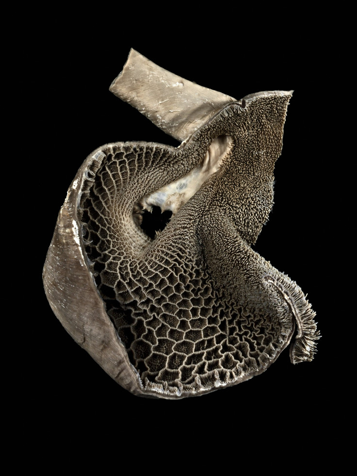
The reticulum, or stomach chamber of a goat. The opening in the center leads to another stomach chamber, the omasum. Goats have four stomach chambers -- the rumen, the reticulum, the omasum and the abomasum.Courtesy of Wellcome Images.Michael Frank, Royal Veterinary College
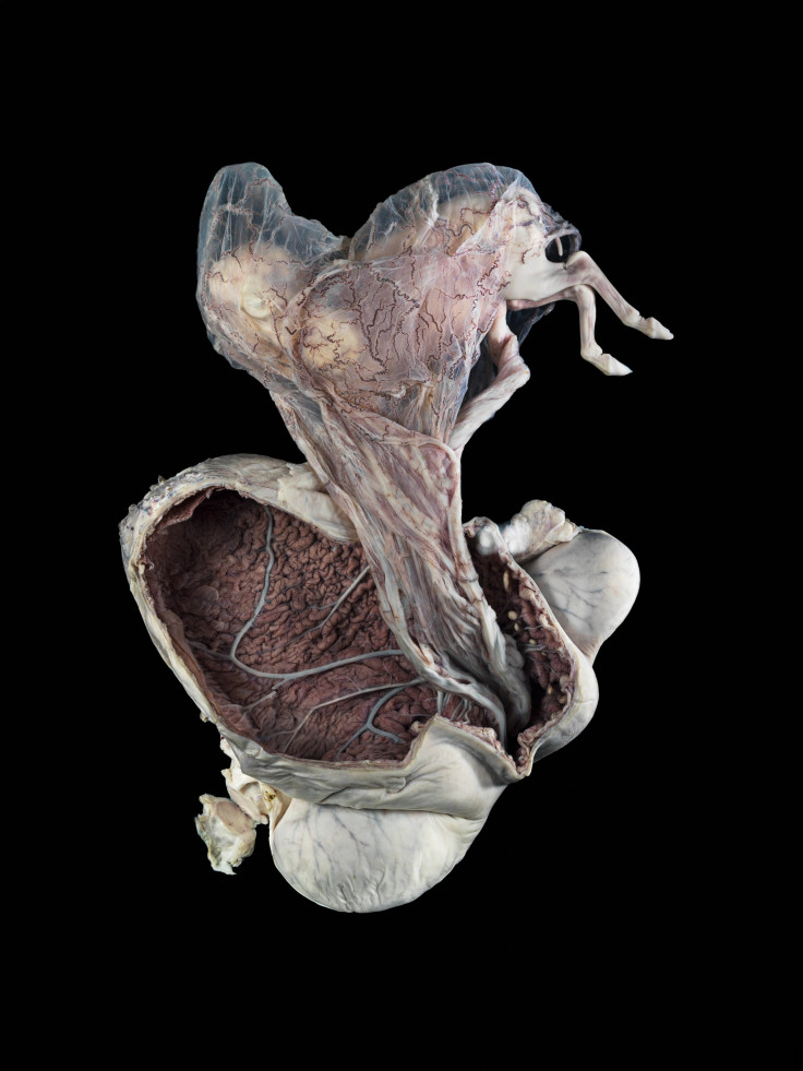
Photograph of a mare's uterus.
Courtesy of Wellcome Images.Michael Frank, Royal Veterinary College
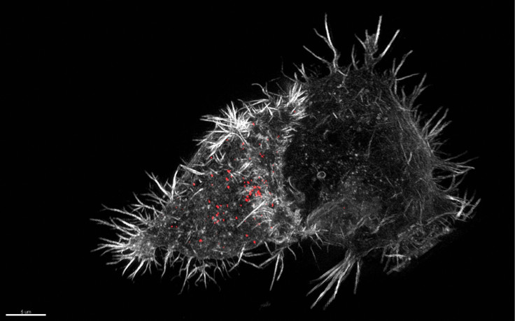
A 3D structured illumination micrograph -- photo taken through a microscope -- of a natural killer cell that's part of our innate immune system. "NK cells produce toxic substances (cytotoxic) which when delivered to a susceptible target cell causes it to self-destruct (apoptosis or programmed cell death)."
Courtesy of Wellcome Images.N. Dieckmann & N. Lawrence, University of Cambridge
A 3D printed rendering of lungs inside the ribcage of a patient with Hodgkin lymphoma cancer.Courtesy of Wellcome Images.Dave Farnham
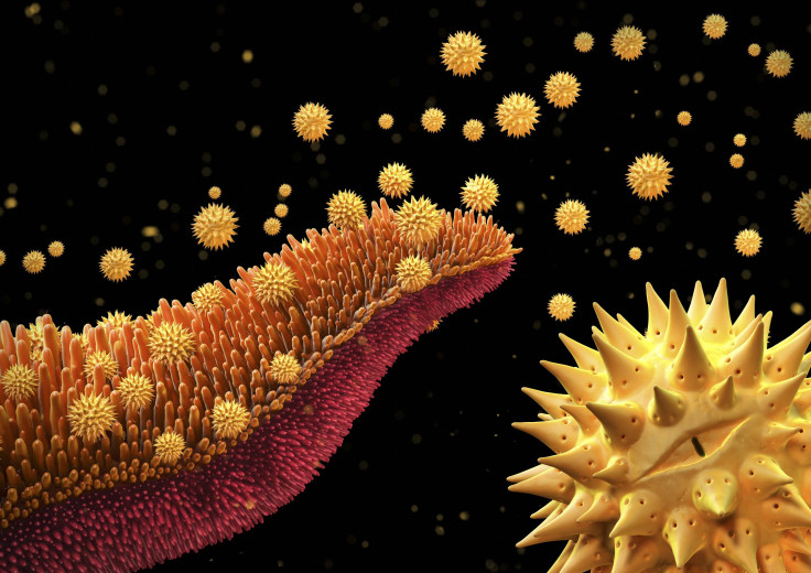
Illustration of pollen grains from a flowering plant.Courtesy of Wellcome Images.Maurizio De Angelis
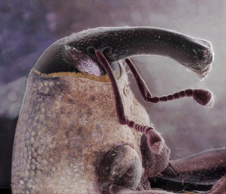
Image of a boll weevil's head. These pests feed and lay eggs in cotton plants.Courtesy of Wellcome Images.Daniel Kariko
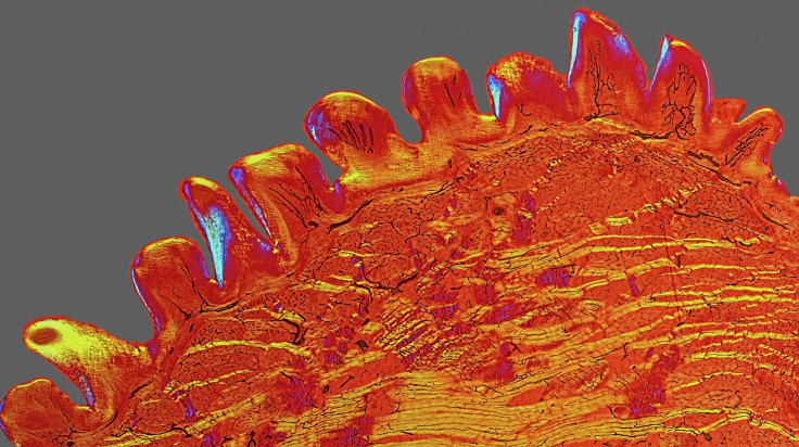
Cross section of a cat's tongue.
Courtesy of Wellcome Images.David Linstead
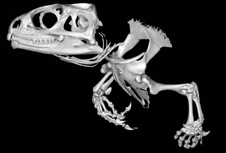
A reconstruction of a tuatara, a rare reptile found only in New Zealand.
Courtesy of Wellcome Images.Sophie Regnault
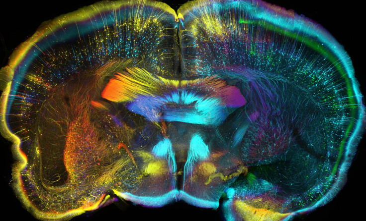
Coronal -- or frontal view -- of a mouse brain. Neurones -- or nerve cells -- are color coded based on depth. At the top is red followed by orange, yellow, purple, blue and green.
Courtesy of Wellcome Images.Luis de la Torre-Ubieta, Geschwind Laboratory, UCLA
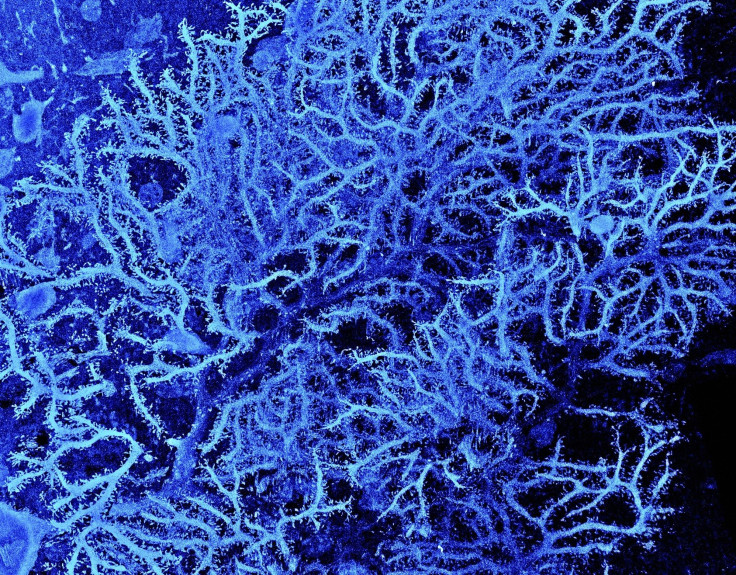
Dendritic tree -- branched projections of neurons -- in the cerebellar cortex of a rat brain. "The cerebellar cortex forms part of the cerebellum, the region of the brain which plays a role in controlling accuracy and coordination of movement."
Courtesy of Wellcome Images.Prof. M. Hausser, Sarah Rieubland & Arnd Roth, UCL
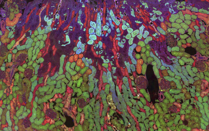
Metabolites inside a mouse kidney. Amino acids aspartate and glutamine are red and blue while the antioxidant glutathione is seen as green in this image. The "resulting image shows how metabolism varies between cells in a single organ. The higher the concentration of the molecule within the cell, the brighter that colour appears."
Courtesy of Wellcome Images.Jefferson R. Brown, Robert E. Marc, Bryan W. Jones, Glen Prusky & Nazia Alam
An astrocyte cell -- a gilal, or non-neuronal cell -- in the brain taking up a carbon nanotube, which could be used asa drug delivery system. The cell is green while the nanotube is brown in this image.
Courtesy of Wellcome Images.Khuloud T. Al-Jamal, Serene Tay & Michael Cicirko
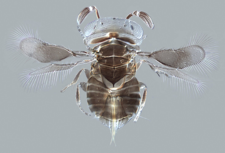
A paraitoid wasp, Wallaceaphytis kikiae, which helps out farmers around the world. "Its close relatives in the genus Aphytis successfully control populations of scale insects (sap-sucking agricultural pests), which attack oranges and other citrus fruits around the world. The female wasps lay their eggs in the scale insects which are then killed by the wasp larvae."Courtesy of Wellcome Images.Andrew Polaszek, Natural History Museum
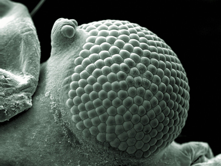
A scanning electron micrograph of a greenfly (aphid) eye.Courtesy of Wellcome Images.Kevin Mackenzie, University of Aberdeen
An elderly woman with Kyphosis.Courtesy of Wellcome Images.Mark Bartley, Cambridge University Hospitals NHS Foundation Trust
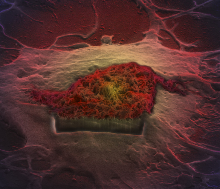
Mouse lungs filled with microparticles carrying drugs (the red/pink spots). ". Current anti-cancer therapies have many toxic side effects so research into other ways of delivering drugs to specific areas of the body are being investigated in order to decrease these unwanted side effects. Here, microparticles were delivered to the lungs using a route similar to drugs administered by an inhaler."
Courtesy of Wellcome Images.Gregory Szeto, Adelaide Tovar, Jeffrey Wyckoff, Koch Institute, copyright MIT
View of a healthy adult, living human brain. The front of head is toward the left of the image and a MRI was used to collect information on the "pathways of white matter in the brain."
Courtesy of Wellcome Images.Dr. Flavio Dell'Acqua
A pediatric sensory unit.Courtesy of Wellcome Images.Geraldine Thompson, CMFT
Old model used in the teaching of Anatomy.Courtesy of Wellcome Images.Anthony Edwards