

Dinosaur Fossils X-Rayed To Study 200 Million-Year-Old Plant-Eating Heterodontosaurus Tucki

Scientists used high-energy X-rays to non-invasively reconstruct the skeletal structure of a 200 million-year-old dinosaur, using fossils discovered in South Africa. The scientists have identified the fossils as the remains of the plant-eating Heterodontosaurus tucki.
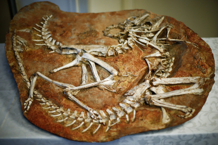
This particular dinosaur was apparently widespread millions of years ago but scientists only had incomplete fossils to analyze prior to this discovery. Scientists at the European Synchrotron Radiation Facility (ESRF) in Grenoble, France, studied the fossils for over five days.
The fossils were discovered by paleontologist Billy de Klerk in South Africa’s Eastern Cape Province in 2005. The dinosaur’s anatomy could not be studied by conventional methods as its skeleton was embedded in hard rock and extracting the brittle bones would have caused a lot of damage. The rock matrix in which the skeletons are embedded reportedly has minerals trapped in it and hence couldn’t be studied under a CT scanner either.
Lead researcher and professor at the Evolutionary Studies Institute in Wits University, Johannesburg, Jonah Choiniere, said in a statement, “There’s still a lot we don’t know about early plant-eating dinosaurs and we need new specimens like this one and new technology like the synchrotron to fill in those gaps.”
The Heterodontosaurus tucki was a small, plant-eating dinosaur with grinding teeth at the back of its jaw and big canines in the front, the scientists found. The scientists studied how the animal ate, moved and breathed using the X-rays produced at ESRF.
“Right away when we open these images we can tell quite a few things about the skull. One of the things is that it's likely a juvenile: the skull bones aren't strongly sutured together. We can also tell that we're really able to reconstruct the skull very, very well,” Choiniere said.
“On the first scans we can see the openings in the skull which are for the balance organs. We can digitally reconstruct the balance organs of the animal and tell how it held its head and how it interacted with its environment. That's the sort of data you just can't get by looking at a skull in 2D. So it's very exciting,” he added.
Great example of collaboration ESRF-SA #synchrotron #paleontology @WitsUniversity @ESI_FossilLab @jonahchoiniere pic.twitter.com/fAVvTh9qNy
— European Synchrotron (@esrfsynchrotron) July 27, 2016
© Copyright IBTimes 2025. All rights reserved.
- MOST POPULAR IN World


