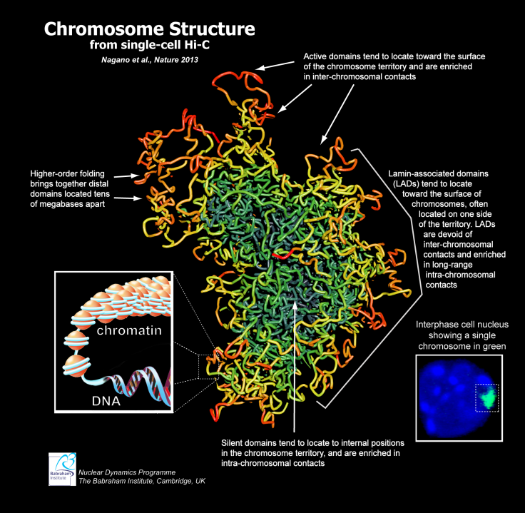
This image shows the chromosome structure from a single-cell Hi-C. The new model of X chromosomes provides a more accurate depiction of the way the chromosomes appears in most cells of a body -- showing that the X-shape generally associated with the them greatly simplifies their structure. Dr. Peter Fraser/Babraham Institute
The well-known X-shape used to visualize chromosomes has been exposed as only a simplified image of chromosomes' true shape.
Scientists at the U.K.'s Babraham Institute have created 3D models that show the true, complex shape of chromosomes and the manner in which DNA folds up within them. Combining research efforts from the University of Cambridge and the Weizmann Institute, the study was published in the journal Nature. The research reveals how the normal X-shape associated with chromosomes only visualizes a specific point in the life of an organism's cells, said Dr. Peter Fraser of the Babraham Institute.
"The image of a chromosome, an X-shaped blob of DNA, is familiar to many but this microscopic portrait of a chromosome actually shows a structure that occurs only transiently in cells -- at a point when they are just about to divide," Fraser said. "The vast majority of cells in an organism have finished dividing and their chromosomes don't look anything like the X-shape. Chromosomes in these cells exist in a very different form and so far it has been impossible to create accurate pictures of their structure."
The new method of visualizing the shape of the chromosomes was created by using the most recent DNA sequencing tools to map out several thousands of molecular measurements for chromosomes in single cells. This data was then compiled, with computers creating 3D images of the chromosomes that reveal their structure and the DNA trail within them.
"These unique images not only show us the structure of the chromosome, but also the path of the DNA in it, allowing us to map specific genes and other important features. Using these 3D models, we have begun to unravel the basic principles of chromosome structure and its role in how our genome functions," Fraser noted.
This new research offers scientists the most accurate visual of how chromosomes -- and the DNA within them -- appear in the body's cells, the Christian Science Monitor reported. By studying the folds and makeup of the chromosomes, more can be learned about the links within chromosomes and how they are connected, Fraser said. This, in turn, could reveal vital information about the regulation of the genome, a vital key to understanding both disease and aging.
Watch the BBSRC's video on the new visualization method below: