

3D-Printed Bacteria ‘Cages’ Allow Scientists To Study Microbe Behavior [PHOTOS]

We’ve seen 3D printers yield everything from prosthetic animal parts to handguns. Now, scientists are 3D printing tiny cages made of protein to house bacterial communities. The microscopic microbial “zoos” replicate conditions where bacteria would normally be found, like in the human lungs and gut.
Scientists at the University of Texas looked at the interactions between bacteria in these 3D-printed environments to better understand what makes some microbes resistant to antibiotics, something health officials have been warning us about for a long time.
"To gain detailed insights into how geometry may influence pathogenicity, we describe a strategy for 3D printing bacterial communities in which physically distinct but chemically interactive populations of defined size, shape, and density can be organized into essentially any arrangement," the researchers wrote.
In other words, scientists from the university used high-precision lasers to print multiple two-dimensional images, using a chip modified from a digital movie projector, onto a layer of flexible gelatin where bacteria were growing. As layers of protein were added to the gelatin, which contain photosensitive molecules that become aroused and bond together after being hit with a laser, they formed a tiny encasing around the bacteria.
Exactly how precise is the laser? “Think about the thickness of a hair on your head, and take 1 percent of that, and then take about a quarter of that,” said Jason Shear, one of the project’s researchers, according to Vice’s Motherboard. “That's about the size of our laser when it's brought to its smallest point.”
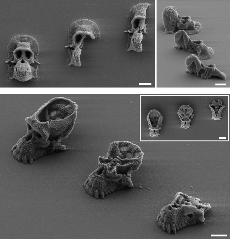
The 3D-printed cages can be made into any shape -- in this case, researchers chose chimpanzee skulls for their miniature microbial zoos. Once inside their cages, the bacteria are kept well-fed with nutrients to make them reproduce.
By trapping the bacteria in these tiny enclosures, researchers were able to pit certain bacteria against others -- like microbial gladiators in a microscopic arena -- and see who came out on top.
Shear and the rest of the team looked specifically at the interaction between Staphylococcus auras, a bacterium that can cause skin infections and often develops a resistance to antibiotics, and Pseudomonas aeruginosa, a bacterium present in cystic fibrosis patients. In the company of Pseudomonas, Staphylococcus became more resistant to antibiotics.
“These are really common bacteria that are often found together in infections, and it makes sense that they would have mechanisms to sense each other,” Shear said. “What the technology allows us to do is put them in conversation with each other, in very precise ways, and see what happens. In this case the Staph sensed the Pseudomonas, and one result was that it became more resistant to the antibiotics.”
As Fox News notes, when Pseudomonas was around, 80 percent of the Staph survived the antibiotic ampicillin. But on its own, only 40 percent of the Staph persisted after an antibiotic attack.
Looking at how bacteria behave en masse is important because bacteria often act different in groups than when they're on their own. In some cases, bacteria clustering can lead to a glue-like substance called biofilm, which clings to surfaces and is more resistant to antibiotics. Learning to counter biofilms is critical to keeping bacterial infections at bay.
Results of the study using 3D-printed bacteria cages were published today in the journal Proceedings of the National Academy of Sciences.
© Copyright IBTimes 2025. All rights reserved.
- MOST POPULAR IN Science


