

New Brain Scan Technique Could Predict Dementia Before Symptoms Are Evident

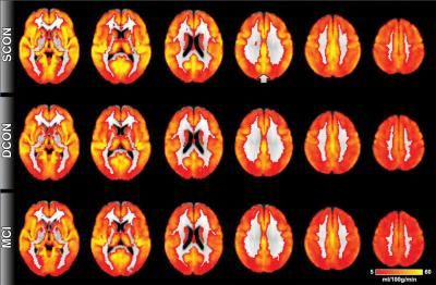
Researchers say a simple technique using magnetic resonance imaging could lead to an early diagnosis of dementia, long before cognitive decline is evident. A study published in the journal Radiology said the MRI technique, arterial spin labeling, or ASL, does not require injection of a contrast agent and could eventually reduce the use of scans exposing patients to radiation.
"ASL MRI is simple to perform, doesn't require special equipment and only adds a few minutes to the exam," study author Dr. Sven Haller of the University of Geneva, in Switzerland, said in a statement.
The study involved 148 healthy seniors and 65 individuals with cognitive impairment. Of the healthy subjects, 73 showed cognitive decline in a follow-up 18 months later, and showed "reduced perfusion at their baseline ASL MRI exams, particularly in the posterior cingulate cortex," the researchers said.
The pattern of reduced perfusion was similar to that of the patients who already had shown signs of cognitive impairment.
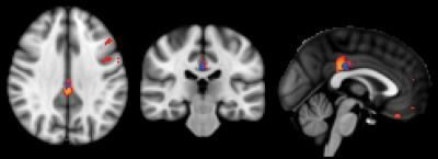
"Less perfusion indicates decreased neural activity," Haller said.
Positron emission tomography, or PET, had previously shown reduced metabolism in the same parts of the brain of Alzheimer's patients as those highlighted by the ASL MRI. Haller said the MRI technique could replace scans for measuring brain metabolism, thus reducing patients' exposure to radiation.
© Copyright IBTimes 2025. All rights reserved.
- MOST POPULAR IN Science


