

2100-Year-Old Egyptian ‘Hawk’ Mummy Turns Out To Be A Child
A group of UK-based researchers has discovered that a 2,100-year-old Egyptian mummy long believed to be that of a hawk actually contains remains of a stillborn human fetus with a rare condition.
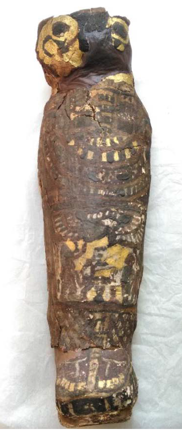
The ancient mummy, kept at the Maidstone Museum, U.K., bore clues of a bird. Its funerary casement had the face of a hawk painted with gilt and was just as big as one would need for a bird. Even the hieroglyphics on it referred to Horus, the falcon-headed deity of the Egyptians.
All these decorations combined with the common practice of animal mummification in ancient Egypt led to the misidentification of the mummy. As a result, it was stored with other animal mummies without conducting CT scans or special attention.
However, the error came to light when the museum decided to scan their resident female mummy as well as a bunch of other animal mummies kept in storage including "EA 493 — Mummified Hawk Ptolemaic Period."
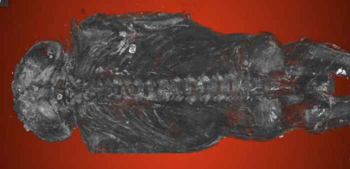
The images of the scan revealed arms crossed over the chest and suggested that there was something else inside — a human or maybe a monkey — but not a bird for sure. They called bioarchaeologist Andrew Nelson from Western University, London, to take a closer look.
Nelson and his interdisciplinary team conducted high-resolution micro-CT scans to virtually unwrap the mummy and found that it contains a severely malformed male human fetus, stillborn between 23 and 28 weeks of gestation. The fetus, as the researchers revealed, suffered from major spinal abnormalities and a rare birth condition called anencephaly, wherein the brain and the skull fail to develop properly.
While the images revealed the mummified fetus had well-formed toes and fingers, the skull bore severe signs of deformities. “The whole top part of his skull isn’t formed,” Nelson said in a statement, noting that the brain of the fetus would not have formed in that scenario. “The arches of the vertebrae of his spine haven’t closed. His earbones are at the back of his head.”
The work, as the researchers said, makes it the second mummified fetus to have been identified with anencephaly as well as the most studied fetal mummy in history.
“The family’s response was to mummify this individual, which was very rare. In ancient Egypt, fetuses tended to be buried in pots, below house floors, in various ways,” Nelson added. “There are only about six or eight known to have been mummified. So this was a very special individual.”
The findings also provide important clues into the diet of the baby’s mother and hint at a lack of foods containing folic acid, which plays a critical role in the development of the neural tube and can lead to anencephaly, if not provided sufficiently. “It would have been a tragic moment for the family to lose their infant and to give birth to a very strange-looking fetus, not a normal-looking fetus at all,” the researcher concluded.
© Copyright IBTimes 2025. All rights reserved.
- MOST POPULAR IN Science


