

Artificial Intelligence Can Help Detect Birth Defects In Ultrasound Images, Study Finds
KEY POINTS
- The study is a pioneer in AI-enabled research in the field of obstetric ultrasonography
- Researchers used deep-learning architecture to detect cystic hygroma
- It's a congenital defect where the lymphatic vascular system develops abnormally
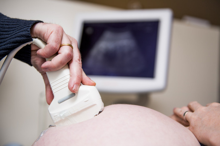
Researchers have used Artificial intelligence (AI) based deep-learning model to diagnose a birth defect in ultrasound images. The experiment bolsters AI’s capabilities to act as an assistive tool for quick and precise reading of ultrasound images.
Dr. Mark Walker and his colleagues at the Ottawa Faculty of Medicine demonstrated the use of deep-learning architecture in the early and reliable diagnosis of cystic hygroma, a congenital defect. The results of the study were published in the peer-reviewed journal, PLOS One.
Cystic hygroma is a condition where the lymphatic vascular system develops abnormally, causing swelling around the head and neck due to the accumulation of fluid. It is a rare disorder that can be life-threatening.
Though the condition can be easily detected in the prenatal stage with an ultrasound, researchers at the University of Ottawa wished to gauge how well AI-driven pattern recognition could do the job. And the results were nothing short of spectacular.
Researchers used around 300 datasets collected at The Ottawa Hospital. The images were analyzed using the DenseNet model to spot cystic hygroma cases with acute sensitivity and specificity in comparison with normal controls. Gradient class activation heat maps—helpful in visualizing pixels in images—were also produced to measure model interpretability.
The results returned a remarkable accuracy of 93%.
"The model was outstanding, even with a small number of training images. And so, potentially, what we demonstrated was in the field of ultrasound we're able to use the same tools for image classification and identification with a high sensitivity and specificity," Walker stated.
The study is a pioneer in AI-enabled research conducted in the field of obstetric ultrasonography, with very few having been done before.
"This particular project is really meant to be the beginning of a large body of work," Walker said. "We've got several papers to follow this one,” he added.
In the future, Walker aims to build an international consortium to upload ultrasound images to remote servers, to expedite diagnosis by physicians in low and middle-income countries with precision. This requires much bigger datasets to be fed to the AI from multiple sources.
© Copyright IBTimes 2026. All rights reserved.
- MOST POPULAR IN Science


