

First 3D-Printed Blood Vessels Bring Scientists A Step Closer To Bioprinting Human Organs

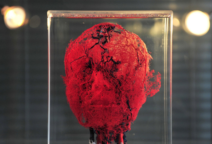
Bioprinting is a growing field in medicine that has scientists well on their way to replicating human organs. But engineering true to life vascular networks has always been a challenge.
Until now.
Scientists in Boston have created the first synthetic blood vessels using 3D-bioprinting techniques. The artificial vessels were used to construct microchannel networks that could be embedded into various types of gels and made into larger organs.
"Engineers have made incredible strides in making complex artificial tissues such as those of the heart, liver and lungs,” Ali Khademhosseini, a biomedical engineer, and director of the Brigham and Women’s Hospital Biomaterials Innovation Research Center, said. "In the future, 3D printing technology may be used to develop transplantable tissues customized to each patient's needs or be used outside the body to develop drugs that are safe and effective.”
Blood vessels transport nutrients and oxygen to tissues and prevent cells from dying. They are also incredibly delicate structures, which makes it difficult to produce them in a lab.
A study published May 6 in the journal Lab on a Chip details how scientists were able to create 3D-print blood vessels from scratch. According to the report, researchers started by making a fiber template from a naturally derived sugar-based molecule called agarose. They enclosed the mold with a gelatin-like substance called hydrogel.
The fibers became the blood vessel channels after the fiber templates were removed.
While the prospect of creating artificial blood vessels is exciting, the question of whether the manmade vessels will perform the duties of the real-life thing remains. As the Voice of Russia noted, the interior walls of human blood vessels contain a special layer of endothelial cells. These cells perform the key functions of vascular biology, such as filtering fluids.
Without that layer of cells, it’s hard to imagine the synthetic vessels being able to support a 3D-printed organ for very long, although scientists did note in their study the successful formation of endothelial monolayers within the channels.
Bioprinting involves using a person’s own body cells to reconstruct an organ. Three-dimensional bioprinting, like other forms of 3D-printing, involves adding layer on top of layer of a material to create a complete structure.
The material used in 3D-bioprinting is made by inserting cells into a biologically safe glue to create a kind of biological paste.
© Copyright IBTimes 2025. All rights reserved.
- MOST POPULAR IN Business


