Facebook And NYU Using AI Research To Make MRI Scans Faster, Simpler
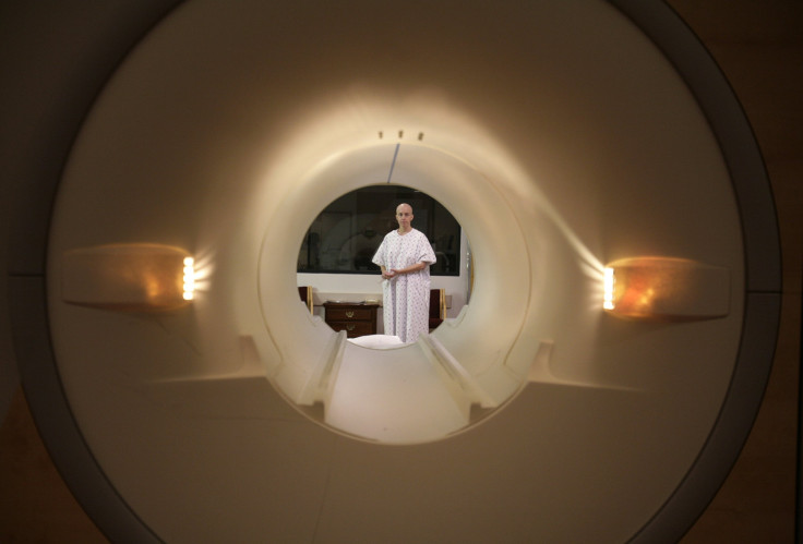
New York University has found an unlikely partner in Facebook on a new project that aims to make MRI scans faster and easier. The project, titled fastMRI, pairs the resources of Facebook’s artificial intelligence research (FAIR) team and radiologists at NYU’s Langone Health to create machines that can produce MRI results with less data.
The two teams have reported teaching an AI using both high and low resolution scans to predict what a final MRI scan will look like using only a fourth of the typical data. This effectively means that fastMRI can deliver accurate results four times faster than current MRI machines, creating a more streamlined experience for patients.
“It’s a major stepping stone to incorporating AI into medical imaging,” FAIR’s Nafissa Yakubova told the Verge.
On Tuesday, the team published a study claiming to prove the accuracy of fastMRI’s AI-extrapolated results in the American Journal of Roentgenology. The study asked radiologists to make assessments of a patient’s knee using traditional MRI scans and fastMRI scans. The results showed that most medical professionals included in the study gave the same assessment for both scans.
“The key word here on which trust can be based is interchangeability,” Dan Sodickson, a radiologist at Langone Health, explained. “We’re not looking at some quantitative metric based on image quality. We’re saying that radiologists make the same diagnoses. They find the same problems. They miss nothing.”
AI upscaling has long been plagued by accuracy issues, usually requiring human eyes to test that the upscaled result matches what was given. The fastMRI team explained that its program is able to avoid these accuracy issues because the low-resolution scans it works with already cover the entire body. The program does not have to guess at missing parts of the image, it just has to bring the scan to a higher resolution.
Also, the program features a check system that allows it to make sure its scan is remaining within the bounds of what traditional MRI machines can producing at regular intervals during the MRI process.
© Copyright IBTimes 2026. All rights reserved.
- MOST POPULAR IN Technology