

First-Ever Color X-Ray Scan Reveals Everything Inside Human Body
We all are pretty familiar with the concept of X-ray scans that help doctors detect minor to major fractures in our bones. The technology has been in use for more than a century, but now, a team of engineers developed a colored X-ray scanner — one that takes ultra-fine photographs of not just your bones but also of your muscles and tissues.
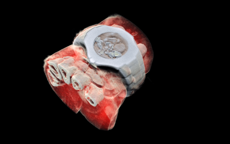
The new MARS spectral x-ray scanner uses an imaging technology developed at the European Organization for Nuclear Research (CERN) and produces sophisticated images showcasing everything lying under your skin marked with different colors.
Till date, X-ray scans have only been about finding and analyzing the anatomy of bones. Doctors pass electromagnetic radiation through a select body part, which gets disturbed due to the presence of bones. The dense material absorbs some of those rays and appears in white in the resulting image, while the remaining rays pass through softer tissues and muscles and appear as solid black.
While this provides a good enough idea of bone structure and condition, there is nothing to learn about the softer part which leaves the rays unimpeded. This is where the new scanner developed by Phil Butler and his son Anthony comes in.
Working for their company MARS Bioimaging, the duo spent more than a decade to come up with a scanner that could provide more information for better medical diagnosis of a patient. They focused on the same scanning mechanism but employed a sophisticated imaging technology developed at CERN called Medipix.
The Medipix chip was initially developed to be used in CERN’s Large Hadron Collider particle accelerator that detected Higgs boson or the “God particle.” In simple terms, the tech detects and counts particles that hit every pixel and enables high-resolution imaging.
MARS Bioimaging leveraged these capabilities to form the third generation of this technology and use it in its full-color, 3D scanner.
The device, as the release described, emitted beams of X-ray, but instead of the conventional way, it measured how the wavelength of the radiation changed after hitting different particles in your body such bone or a tissue. As this happens, a sophisticated algorithm uses that information to produce a 3D image with different colors representing different materials scanned. This enables easy differentiation between bones, fats, muscles, calcium, disease markers or all other tissues present in the body.
“This technology sets the machine apart diagnostically because its small pixels and accurate energy resolution mean that this new imaging tool is able to get images that no other imaging tool can achieve,” Phil Butler said in a release.
The working of the scanner has been proved, but it still has to go through a number of trials and regulatory checks before being used in modern-day clinics. However, whenever that happens, doctors would be able to use it for accurate diagnosis of cancer or arthritis than currently possible.
“In all of these studies, promising early results suggest that when spectral imaging is routinely used in clinics it will enable more accurate diagnosis and personalization of treatment,” Anthony Butler concluded in the statement.
© Copyright IBTimes 2025. All rights reserved.
- MOST POPULAR IN Technology


